Microscopy workshop
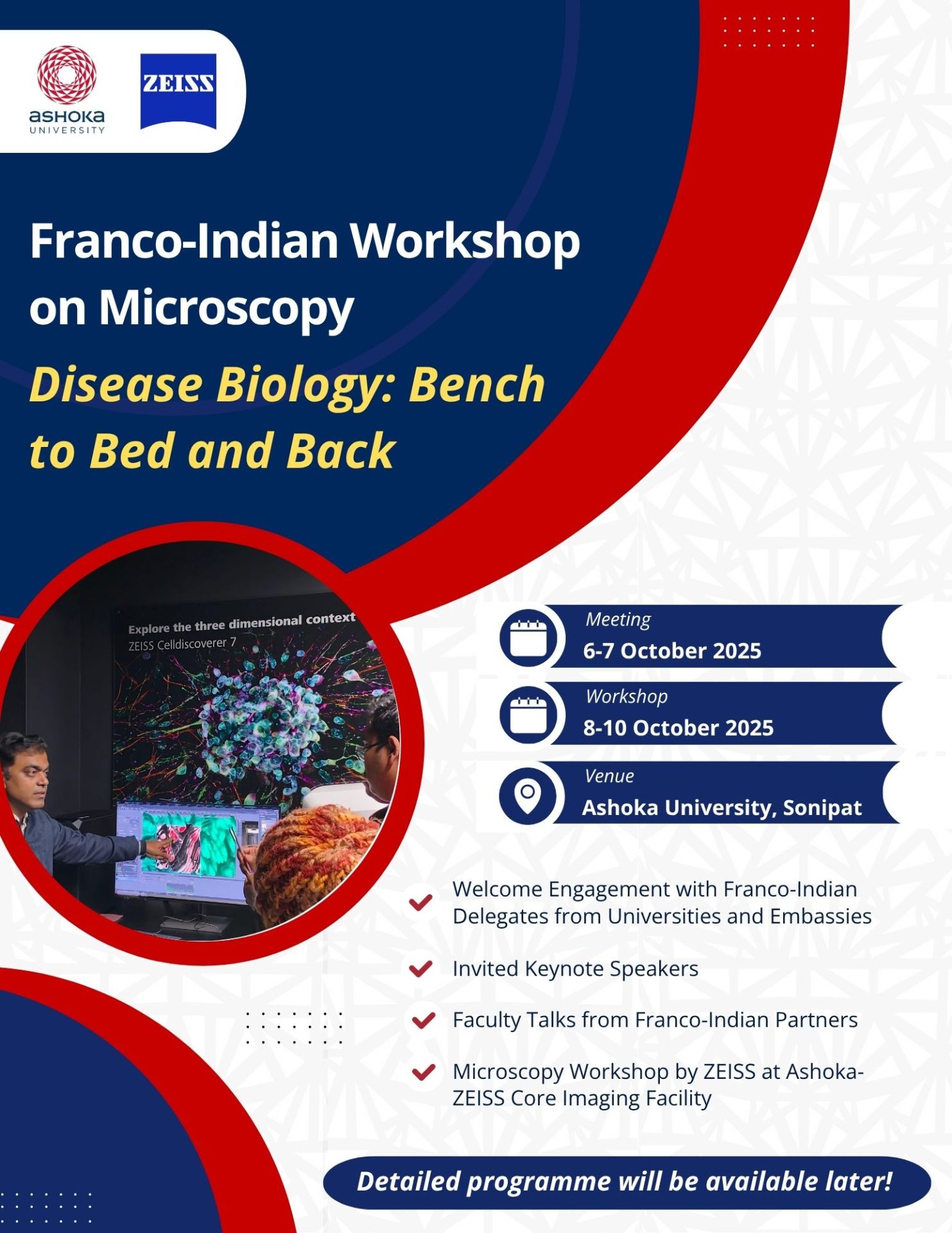
Franco-Indian Workshop on Microscopy
Disease Biology: Bench to Bed and Back
Ashoka University, Sonepat – 6–10 October 2025
The Franco-Indian Workshop on Microscopy was conceived as a platform to foster scientific
dialogue, interdisciplinary exchange, and hands-on capacity-building in advanced imaging for
biomedical research. Its primary objectives are to:
• Strengthen collaboration between French and Indian institutions under the umbrella of the
Franco-Indian Campus in Life Sciences for Health.
• Showcase cutting-edge research in disease biology—from molecular mechanisms to
translational applications.
• Train a new generation of researchers in state-of-the-art microscopy techniques through
immersive, practical sessions.
• Promote cross-fertilisation of ideas linking biological discovery, technological innovation, and
clinical impact.
Organised by Ashoka Global Research Alliances in partnership with the Franco-Indian Campus in Life Sciences for Health, this workshop unites researchers and students from both nations around a shared ambition: advancing disease biology through the lens of cutting-edge microscopy. The event serves as a platform to deepen Franco-Indian scientific cooperation, promote interdisciplinary exchange, and encourage the circulation of expertise across research and training.
The workshop is designed to strengthen the bridge between fundamental biology and its clinical applications. It seeks to nurture collaborative research and to provide young scientists with direct exposure to the most advanced imaging technologies available today. Through shared experimentation and dialogue, participants explore how microscopy connects molecular mechanisms to complex physiological and pathological processes, enabling a true “bench-to-bed and back” approach that integrates laboratory discovery with clinical insight.
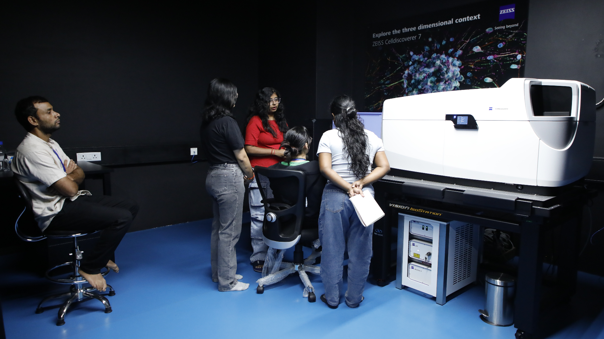
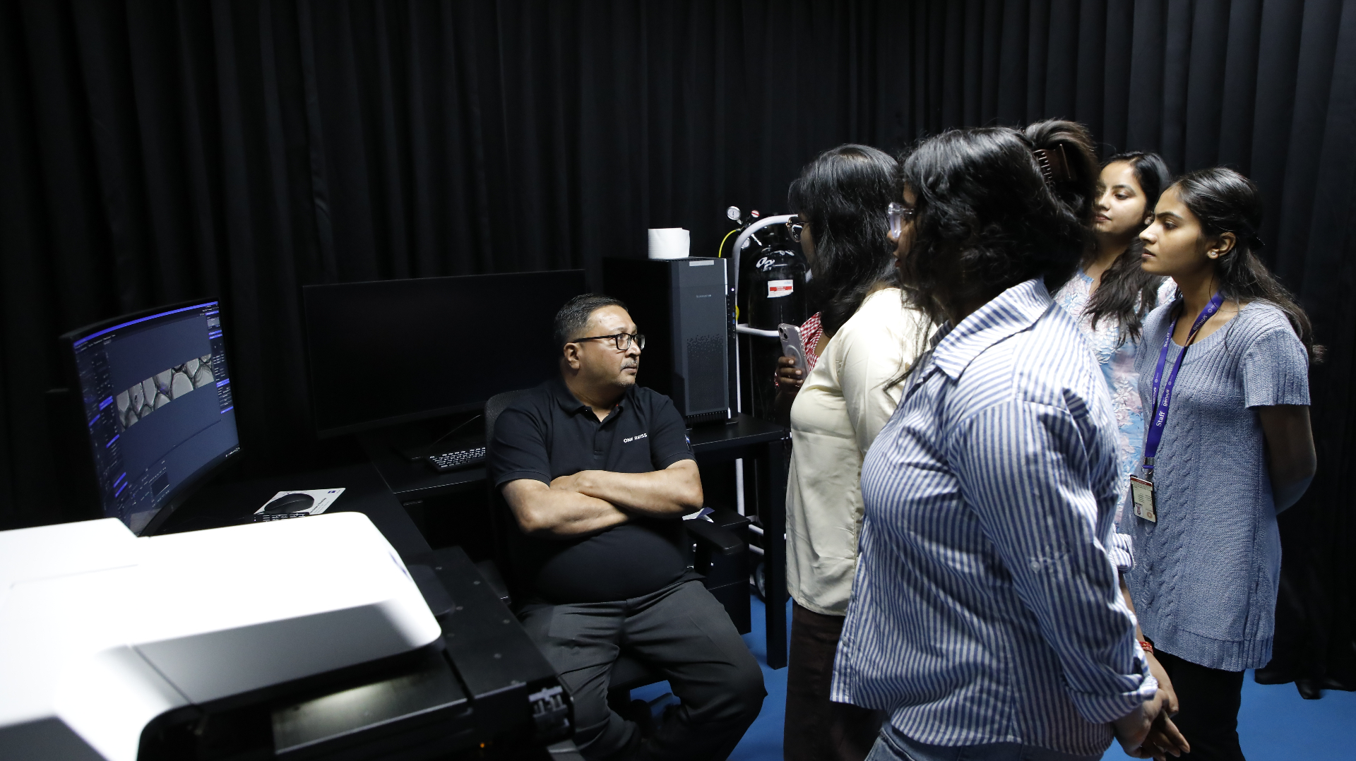
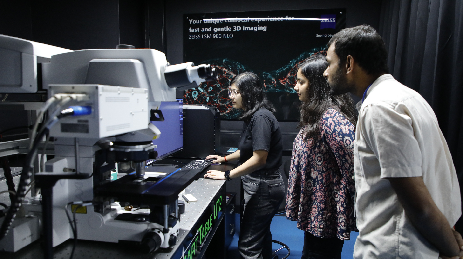
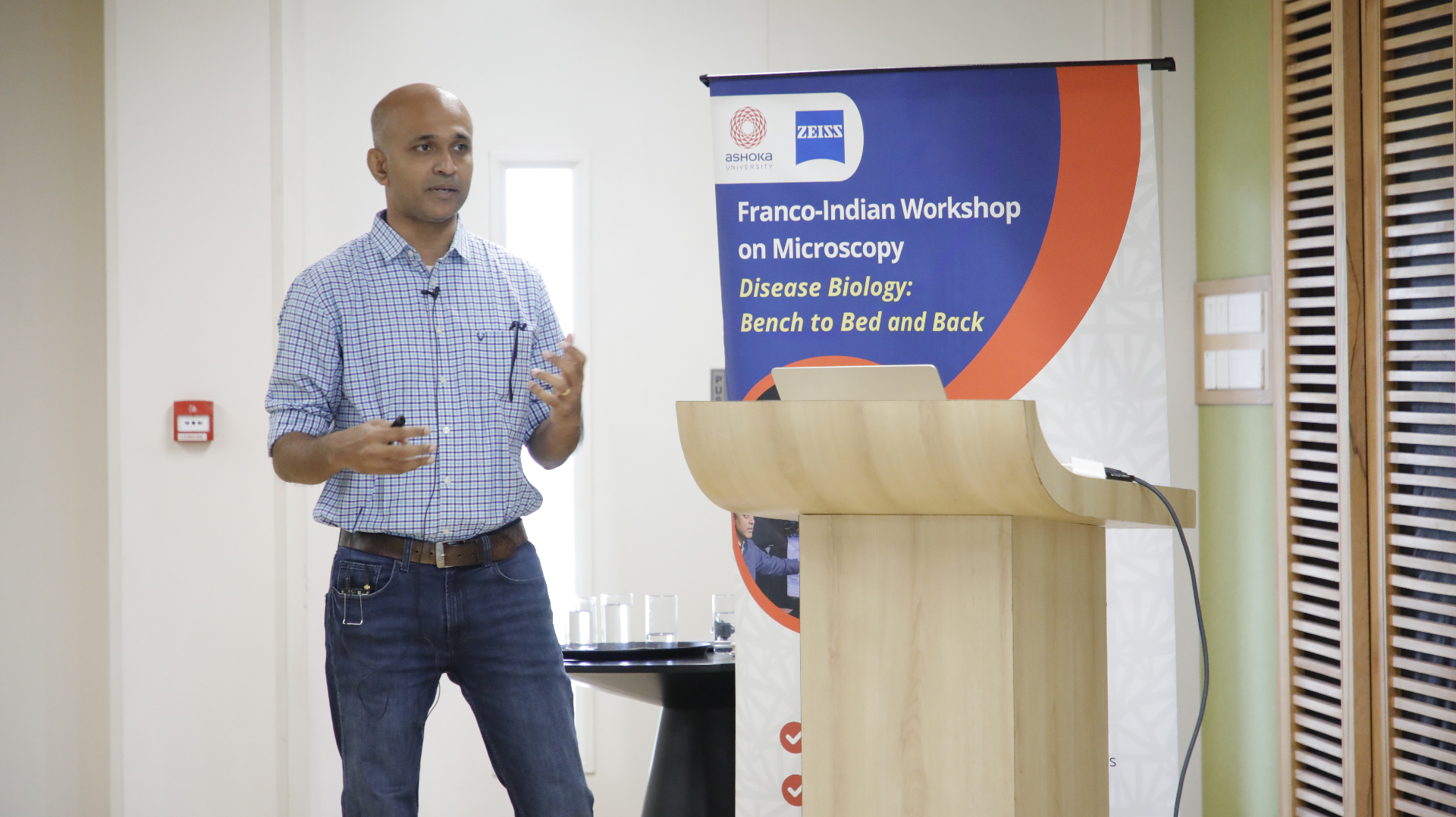
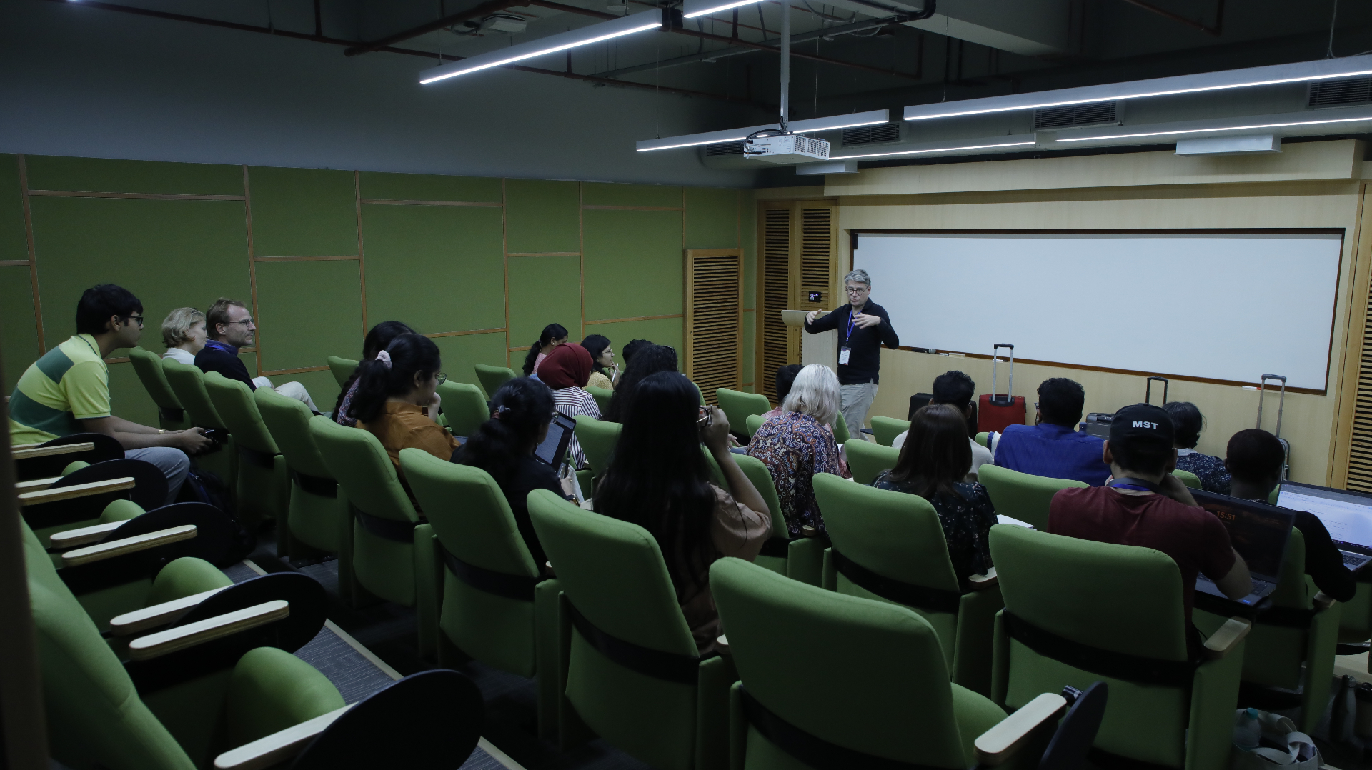
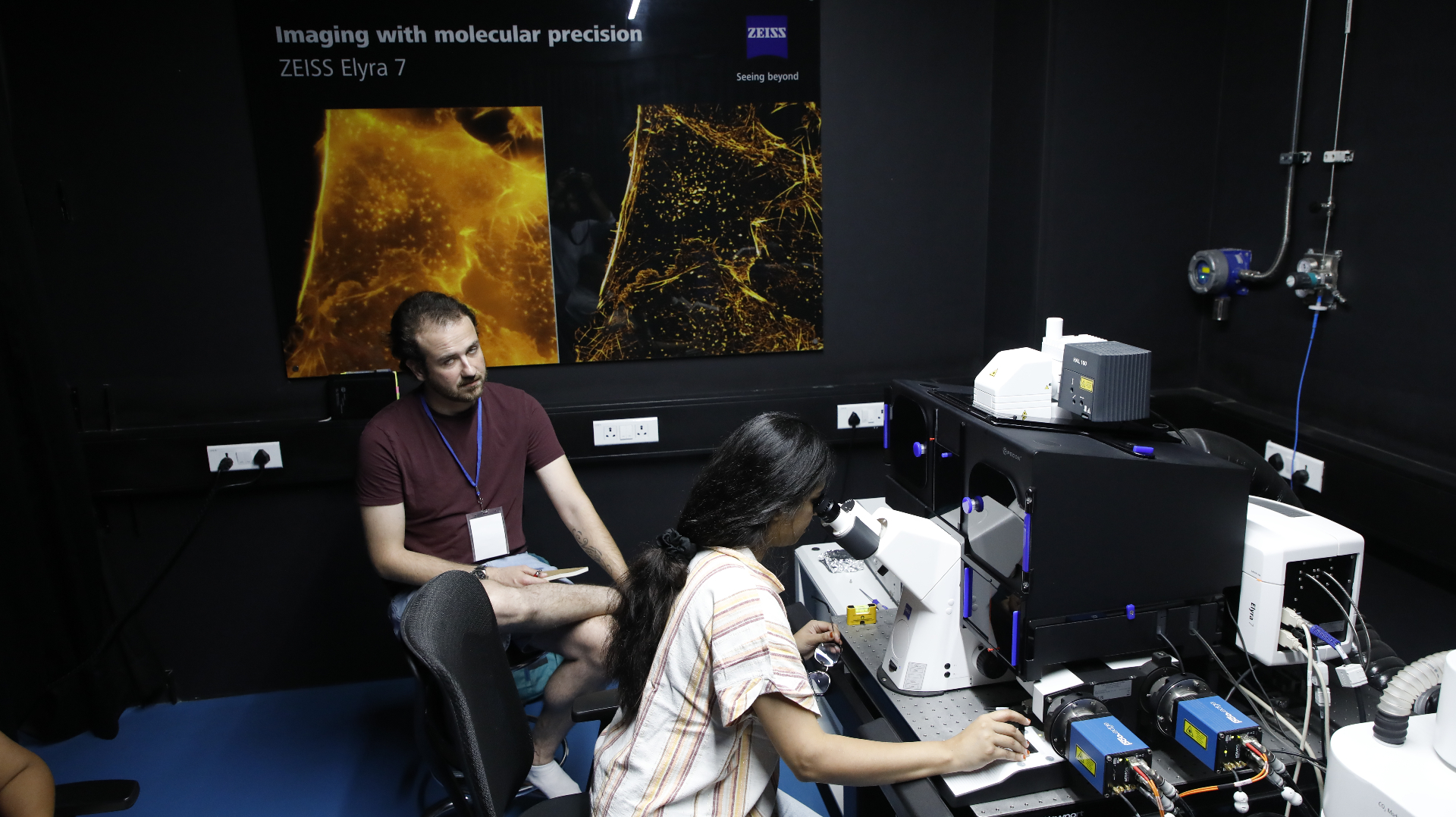
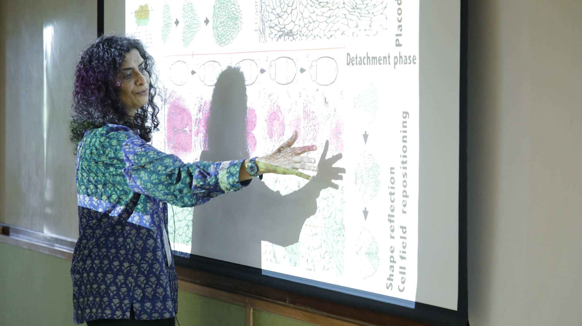
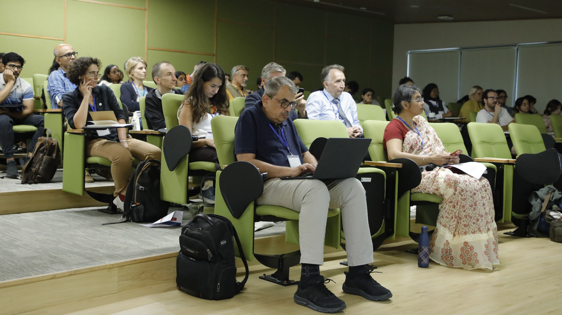
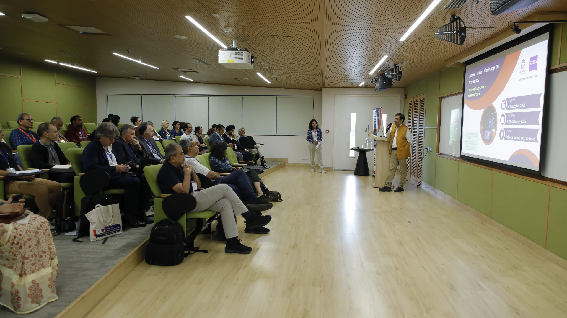
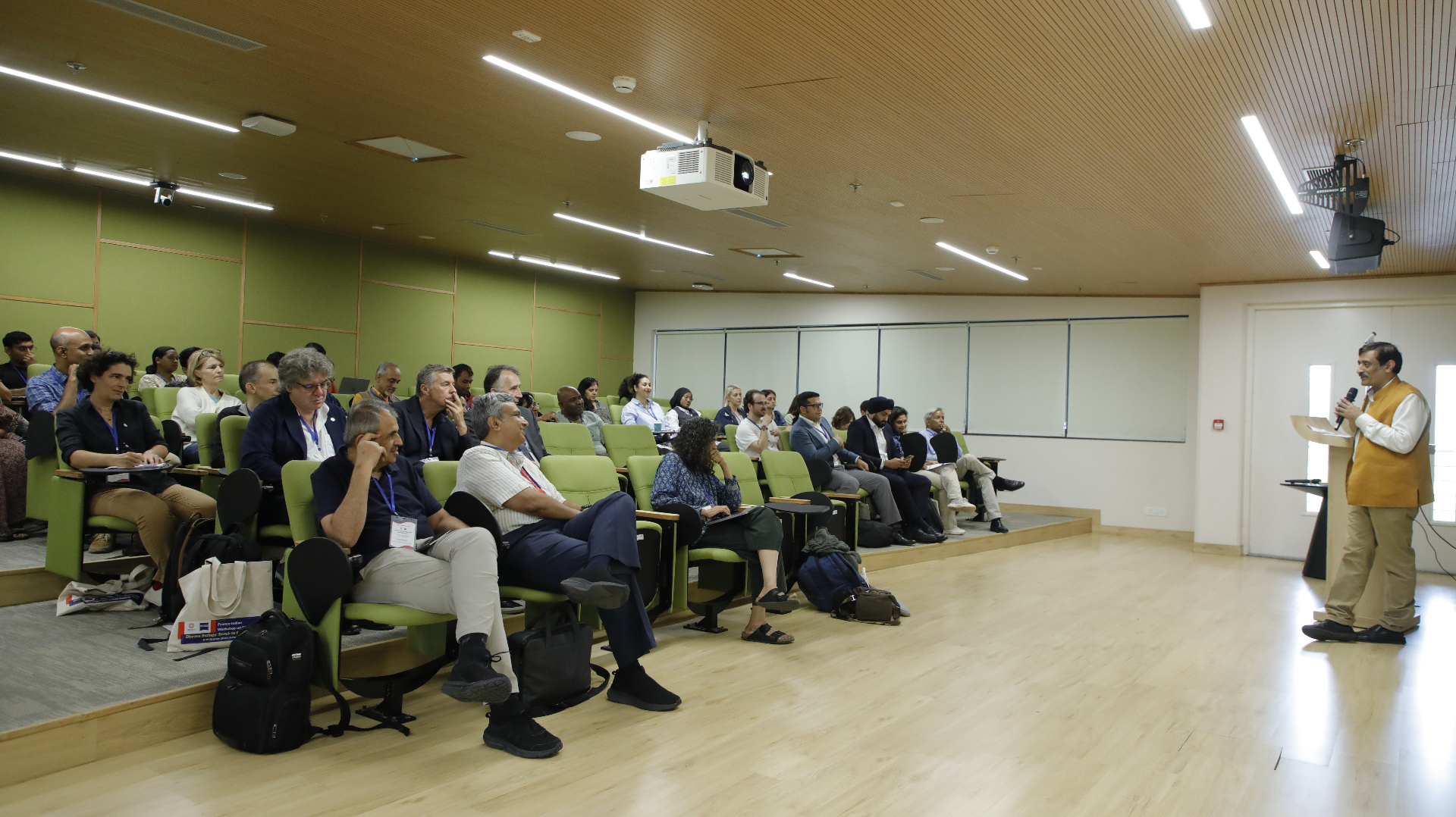
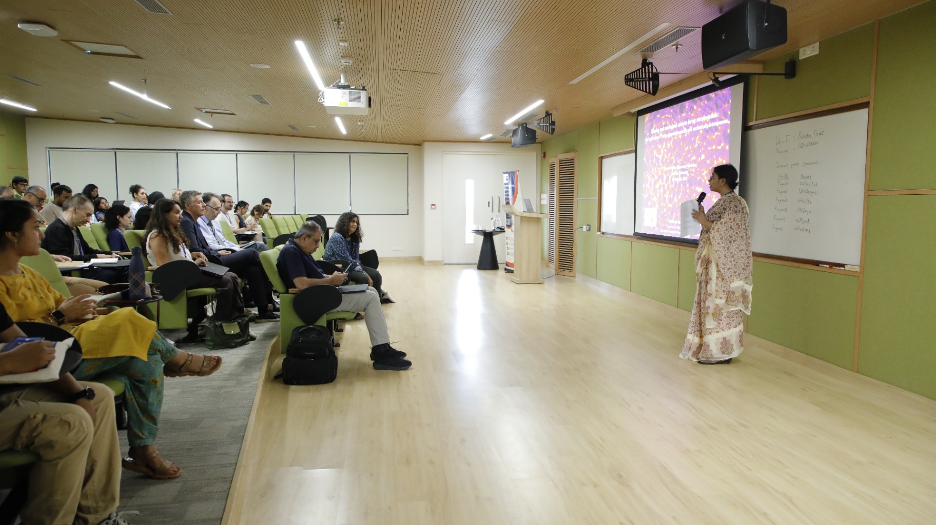
Over the course of five days, the programme combines academic presentations with hands-on sessions. The opening segment at Ashoka University features scientific discussions led by faculty from the four FIC consortia, who present recent advances in cellular imaging, disease modelling, and computational analysis. The latter part of the week unfolds in the Ashoka–Zeiss Core Imaging Facility, where participants work in small groups under expert supervision. This immersive format allows for direct engagement with high-resolution imaging platforms, experimental design, and data interpretation in a collaborative environment.
The scientific focus of the workshop reflects the diversity and richness of microscopy as a research tool. Discussions encompass the dynamics of cells and molecules, the fine architecture of neuronal networks, and the imaging of cancer and metabolic disorders. Sessions also delve into the visualisation of developmental processes and morphogenesis, while highlighting the emerging role of artificial intelligence in image analysis and diagnostic innovation. Technological advances in organic dyes, expansion microscopy, and the study of phase separation are presented as part of a broader effort to push the frontiers of biomedical imaging.
By bringing together expertise, technology, and vision, this workshop contributes to the creation of a vibrant Franco-Indian community of practice in microscopy and imaging sciences. It opens new pathways for joint research initiatives, fosters long-term academic exchanges, and reinforces the visibility of the Franco-Indian Campus in Life Sciences for Health as a model of Indo-Pacific scientific collaboration grounded in excellence, innovation, and mutual trust.
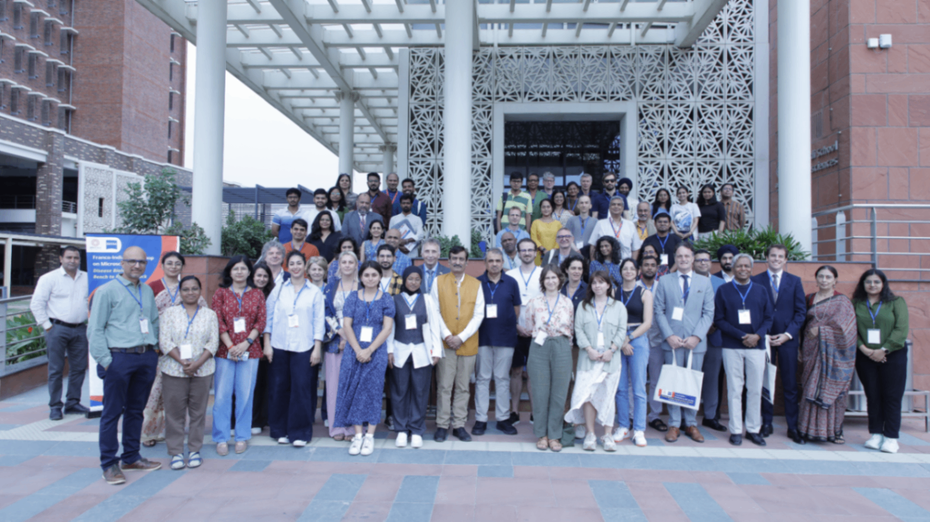
Event Images